
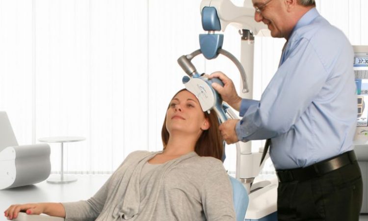
La stimulation magnétique transcrânienne (TMS) est une technique de neuromodulation utilisée depuis 30 ans. Cette méthode utilise un champ magnétique alternatif pour induire une excitation électrique dans une zone précise du cortex, via l’induction électromagnétique. Un stimulateur génère un courant pulsé, connecté à une bobine magnétique placée contre le crâne. Le stimulateur contient un condensateur qui produit un courant de tension allant jusqu’à 3000 V et une intensité atteignant 20 000 A. Cela entraîne la dépolarisation de la membrane cellulaire des neurones et l’activation du cortex directement situé sous la zone stimulée. Cette excitation peut se propager à travers les synapses et les neurones voisins vers les structures du cerveau auxquelles cette zone corticale est reliée. La TMS permet ainsi de stimuler le cerveau de manière électrique, sans utiliser d’électrodes.
La stimulation du cortex moteur dans la région responsable du mouvement des doigts ou des orteils est réalisée afin de vérifier que, chez un patient inconscient, il n’y a pas de rupture de la moelle épinière (pour voir si l’impulsion provenant du cortex moteur peut traverser la voie cortico-spinale intacte). Un seul stimulus appliqué à une zone appropriée du cortex peut également perturber les interactions neuronales en cours dans cette zone. Ainsi, par exemple, il est possible de démontrer l’implication d’une région corticale donnée dans certains processus perceptifs.
La TMS permet également de cartographier les connexions au sein du cerveau : les effets peuvent être observés non seulement dans la structure stimulée, mais aussi dans les régions auxquelles cette structure est reliée. Elle permet également de stimuler ou d’inhiber une région spécifique du cortex, grâce à une série d’impulsions – appelée rTMS (TMS répétitive). Les impulsions appliquées à une fréquence d’environ 4 Hz ont un effet stimulant, qui peut durer de quelques minutes à plusieurs dizaines de minutes après la fin de la stimulation. Dans le cas de la stimulation inhibitrice, on crée ce qu’on appelle une “lésion virtuelle”, c’est-à-dire un blocage temporaire de l’activité d’une région choisie du cortex. La première démonstration de cette méthode a été l’inhibition temporaire du langage en créant une “lésion virtuelle” de la zone de Broca chez des volontaires sains. Dans une étude célèbre sur la plasticité cérébrale, des sujets ont été bandés les yeux pendant une semaine et ont appris à lire le braille. Après une semaine, il s’est avéré que les stimuli tactiles (les lettres en relief du braille) n’étaient pas traités par le cortex somatosensoriel, mais par le cortex visuel – une réorganisation plastique du cerveau avait eu lieu. Cela a été prouvé en interrompant l’activité du cortex visuel avec la TMS : les sujets étaient alors incapables de distinguer les lettres en braille.
À Quoi Sert la TMS en Thérapie ?
Depuis des années, les spécialistes cherchent des applications thérapeutiques pour la TMS. De grands espoirs ont été mis dans l’ajout de la TMS à la neuro-rééducation après un AVC. La version la plus fréquemment étudiée est la suppression, par TMS, de l’inhibition interhémisphérique. Dans les zones motrices et sensorielles du cortex cérébral, l’effet d’un hémisphère sur l’autre est inhibiteur. On pensait que, par exemple, en cas de lésion unilatérale du cortex moteur suite à un AVC, les effets inhibiteurs de l’hémisphère intact pourraient entraver les modifications plastiques que la rééducation visait à déclencher dans les zones intactes autour du site de l’AVC. Certaines tentatives ont amélioré le processus de rééducation, mais de nombreux échecs ont également été observés. Des essais ont aussi été menés pour traiter l’aphasie (avec peu d’effet) et la négligence spatiale (avec des résultats plus prometteurs).
La réduction de la douleur chronique a fait l’objet de plusieurs expériences dans lesquelles la TMS a été utilisée pour diminuer la douleur neuropathique. Malgré des résultats positifs dans de petits groupes expérimentaux, les méta-analyses n’ont pas montré d’efficacité clinique.
